Speed, Precision & Specialized Cellular Profiling
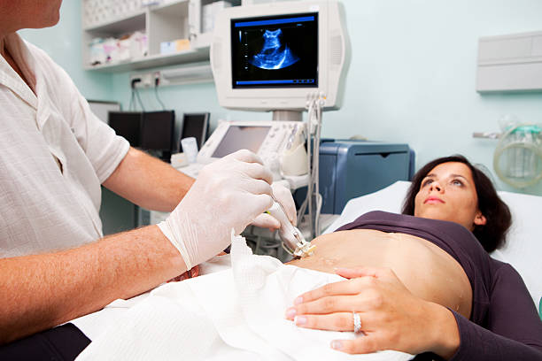
The pathology services at Gauri Shukla Hospital provide immediate, comprehensive tissue evaluation and expert microscopic verification for suspicious masses, ulcers, and chronic inflammatory mutations.
Our central diagnostic lab prioritizes patient safety, absolute sample fixation integrity, and highly accurate laboratory reporting in every cellular scanning scenario. The processing pipeline begins with rapid clinical triage labeling and specialized formalin container registration, ensuring priority-based targeted sample tracking without cross-contamination or processing delays. Our trained laboratory technicians, specialized histopathologists, and clinical medical staff follow strict international verification standards to balance microtome slicing calibrations, analyze delicate cellular architectures, and safely issue actionable medical data.
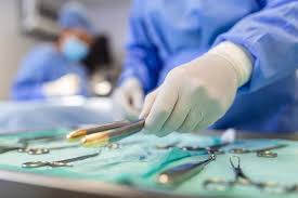
Equipped for High-Precision Tissue & Micro-Slicing Assay
Our specialized diagnostic laboratory is fully integrated with computerized tissue embedding stations and high-resolution optical microscopes to process all biopsies safely and effectively.
- Advanced Rotary Microtomes for Ultra-Thin Micron Sectioning
- Automated Vacuum Tissue Processors and Paraffin Embedding Blocks
- High-Definition Trinocular Microscopes with Digital Image Capture
- Specialized Cryostat Stations for Rapid Intraoperative Frozen Sections
- Advanced Immunohistochemistry (IHC) Biomarker Staining Grids
- Direct Digital Integration with 24/7 Surgical Theaters and Ward Terminals
Biopsy & Pathology Services Offered
The Tissue Processing & Analysis Journey
Sample fixation sterility, meticulous paraffin wax embedding, and expert microscopic tracking form the framework of our diagnostics path.
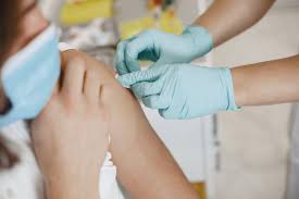
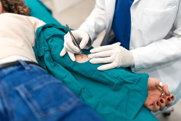
Extraction & Chemical Fixation
Procurement of target cells and prompt immersion in balanced formalin solutions to lock cellular structures safely.
Gross Examination & Logging
Meticulous physical measuring, color description profiling, and secure generation of unique electronic block barcodes.
Paraffin Embedding
Following standardized clinical protocols to dehydrate tissues and encase specimens into solid paraffin wax casting blocks.
Microtome Section Slicing
Cutting precise, paper-thin tissue sheets measuring 3-5 microns and mounting sections safely onto diagnostic glass slides.
Staining & Microscopy
Continuous monitoring of chemical H&E setups and professional microscopic screening of cellular architectures by a pathologist.
Verified Expert Report
Coordinated validation of tumor classifications, grading metrics, or clearance margins for secure integration into health portals.
Emergency Lab & Biopsy Support Helpline: +91 72800 09000
Our highly responsive pathology lab is active 24/7 to receive urgent surgical biopsy specimens, execute rapid intraoperative frozen section protocols, or check tumor margin integrity with advanced analytical support. Do not delay under critical diagnostic uncertainty—quick response is our priority.
Advanced Readiness & Histopathological Precision
Our biopsy testing workflows are planned meticulously to guarantee complete sample safety, eliminate misdiagnosis anomalies, and ensure clear clinical verification windows.
- 24/7 absolute tissue processing access with senior clinical histopathologists on active call
- Fully integrated modular laboratories featuring advanced automated staining arrays and crisp optical imaging lines
- Standardized pre-analytical tracking checklists for rapid oncological staging, tumor margin mappings, and deep node evaluations
- Highly supportive laboratory technical team prioritizing zero-contamination tracking and total patient privacy safety